Skip to content

Vascular Ultrasound
- Home
- Vascular Ultrasound
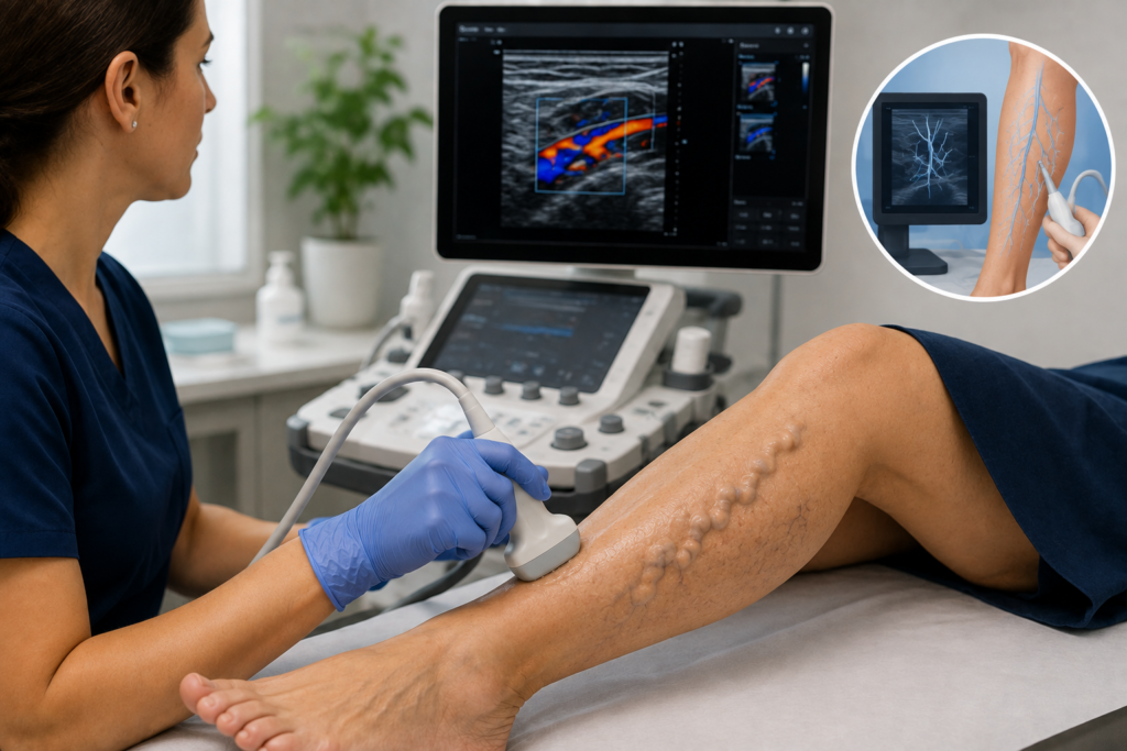
Vascular Ultrasound & Mapping in Melbourne
Venous ultrasound and vein mapping are diagnostic imaging methods used to examine veins, assess blood flow and identify abnormalities that may contribute to varicose veins and other venous conditions.
A venous duplex ultrasound combines detailed images of the veins with Doppler technology to assess blood-flow direction and valve function. This can help identify venous reflux, where blood flows backwards through veins because the valves are not functioning effectively.
What Is Venous Ultrasound and Vein Mapping?
Venous ultrasound is a non-invasive medical imaging examination that uses high-frequency sound waves to produce images of veins and assess blood flow.
When combined with Doppler techniques, the examination is called venous duplex ultrasound. It helps clinicians evaluate vein structure and blood-flow patterns, including whether blood is flowing in the expected direction.
Vein mapping is a specialised application of duplex ultrasound that documents relevant vein anatomy and function. It is commonly used when investigating varicose veins or planning treatment for venous insufficiency.
How Does Venous Duplex Ultrasound Work?
The examination combines two techniques:
B-mode ultrasound: Produces images of vein structures, allowing the examiner to assess vein location, size and anatomy.
Doppler ultrasound: Evaluates blood movement, including its direction and flow characteristics.
Together, these techniques help identify abnormal blood flow and assess selected superficial and deep veins.
What Is the Difference Between Venous and Arterial Ultrasound?
A clinician may recommend a vein ultrasound when symptoms, medical history or examination findings suggest an underlying venous circulation problem.
Common reasons for considering venous ultrasound include:
Varicose Veins
Varicose veins are enlarged, often twisted superficial veins that may be visible beneath the skin. Duplex ultrasound can help determine whether visible varicose veins are associated with abnormal valve function or reflux in larger superficial veins.
Leg Pain, Aching or Heaviness
Some patients with venous insufficiency experience leg aching, heaviness or discomfort, particularly after prolonged standing. Ultrasound may be considered when symptoms and clinical findings suggest a venous cause. These symptoms can also occur with other medical conditions, so assessment should not assume that veins are responsible.
Leg Swelling
Persistent or recurrent leg swelling can have several causes. Where venous disease is suspected, ultrasound may help investigate relevant vein abnormalities. Additional investigations may be necessary depending on the patient’s symptoms and medical history.
Skin Changes or Venous Ulcers
Changes such as pigmentation, eczema-like irritation or wounds around the lower legs may sometimes be associated with chronic venous insufficiency. Venous ultrasound can assist in assessing underlying reflux where clinically indicated.
Recurrent Varicose Veins
Patients who develop new or recurrent varicose veins after previous procedures may require ultrasound assessment to examine altered vein anatomy and identify potential sources of reflux.
Before Certain Vein Treatments
Ultrasound mapping may be used to investigate vein anatomy and plan appropriate treatment when an intervention is being considered. Not everyone with spider veins or other small visible surface veins requires duplex ultrasound. The need for scanning depends on individual clinical findings.
Important: Sudden swelling or pain in one leg can be a sign of deep vein thrombosis (DVT) and requires prompt medical assessment. A routine varicose vein mapping appointment is not a substitute for urgent DVT investigation.
What Can Duplex Ultrasound and Vein Mapping Identify?
Venous duplex ultrasound helps clinicians examine the anatomy and function of relevant veins beneath the skin.
The findings may include:
Venous Reflux and Valve Function
Healthy veins contain valves that help blood move towards the heart.vWhen these valves do not close effectively, blood may flow backwards. This is called venous reflux and may contribute to varicose veins and chronic venous insufficiency.vDoppler ultrasound can assess the direction of blood flow and help identify veins affected by reflux.
Superficial and Deep Vein Anatomy
Vein mapping may examine relevant superficial veins, including the great saphenous vein and small saphenous vein, along with their connections to other veins. Depending on the clinical purpose, the examination may also assess aspects of the deep venous system.
Abnormal Vein Connections
Some visible varicose veins are connected to larger veins with underlying reflux. Mapping can help identify these connections and provide information that may be useful in developing an appropriate management plan.
Vein Diameter and Reflux Distribution
Where relevant, the examiner may record vein measurements, reflux patterns and the location of abnormal blood flow. These findings are considered alongside symptoms, clinical examination and other medical information.
Venous Reflux Studies vs DVT Ultrasound
A venous reflux study and an ultrasound examination for suspected DVT have different primary purposes. A reflux study assesses vein function and abnormal blood-flow direction. A dedicated DVT ultrasound investigates possible blood clots using a protocol appropriate to that clinical question. The examinations may overlap in some components, but one should not be assumed to replace the other.
How Is a Venous Ultrasound Performed?
A venous duplex ultrasound is usually performed externally using a handheld ultrasound probe placed against the skin. The exact procedure depends on the reason for the examination, the veins being assessed and the patient’s medical circumstances.
A typical varicose vein assessment may involve:
Reviewing your symptoms and history: The clinician or ultrasound professional confirms the purpose of the investigation and any relevant medical information.
Positioning for the examination: You may be asked to stand, sit or lie down so that the relevant veins can be examined.
Applying ultrasound gel: A water-based gel is applied to the skin to help transmit sound waves.
Scanning the veins: A handheld probe is moved over the leg to produce images of the veins and assess blood flow.
Testing blood-flow direction: The examiner may use gentle calf compression or other manoeuvres to assess venous valve function.
Recording findings: Relevant vein measurements, blood-flow patterns and abnormalities are documented for clinical interpretation.
The examination may involve scanning different areas of the leg, including the thigh, calf and behind the knee. The amount of time required depends on the type and extent of the examination. The ultrasound professional can explain what is being assessed and advise you if any positioning changes are needed.
Preparing for Your Ultrasound Examination
Preparation for a standard leg vein ultrasound is generally straightforward, although instructions may vary depending on the requested examination.
Before your appointment, you may be advised to:
Wear loose, comfortable clothing that allows access to your legs.
Bring any referral or appointment documentation requested by the clinic.
Provide previous ultrasound reports or relevant vein treatment records.
Bring a current list of medications where relevant.
Inform the clinic if you have difficulty standing or remaining in one position.
Follow any instructions about compression stockings or other garments.
Fasting is generally not required for routine leg venous ultrasound. Other vascular examinations can have different preparation requirements.
Do not stop prescribed medication or change your usual medical care unless instructed by your treating clinician. If you are unsure about preparation, contact Vein Care before your appointment.
Is Venous Ultrasound Safe or Uncomfortable?
Venous ultrasound is a non-invasive diagnostic examination that uses sound waves rather than ionising radiation. For a standard external leg vein examination, it does not usually require needles, surgical incisions or contrast injections.
Most patients tolerate the examination well. However, temporary discomfort may occur if:
The probe applies pressure over an already tender area.
The examiner compresses the calf to assess blood flow.
You need to stand or maintain a particular position.
You have painful or swollen veins.
Tell the examiner if you experience pain, dizziness or difficulty remaining in the requested position.
Understanding Your Results and Next Steps
After a venous duplex ultrasound, the findings are assessed in relation to the reason the examination was requested. The results may identify venous reflux, document relevant vein anatomy or show that the symptoms require consideration of another possible cause.
What Happens If Venous Reflux Is Found?
Venous reflux does not automatically mean that an invasive treatment is necessary.
A clinician considers factors such as:
Your symptoms and how they affect daily activities
The location and extent of abnormal reflux
The size and pattern of affected veins
Skin changes or other complications
Previous vein treatment
Relevant medical conditions
Your preferences and treatment goals
Depending on the findings, management may include monitoring, conservative measures, further investigation or consideration of an appropriate vein procedure.
What If the Ultrasound Does Not Explain My Symptoms?
Leg symptoms can have causes other than venous disease. If ultrasound findings do not explain the symptoms, the clinician may recommend further medical assessment or referral where appropriate. Ultrasound provides diagnostic information. It does not independently determine a diagnosis or the need for treatment without clinical interpretation.
How Ultrasound Supports Vein Treatment Planning
Duplex ultrasound plays an important role in assessing many patients with clinically significant varicose veins. Visible varicose veins can be associated with reflux in larger superficial veins that are not fully apparent during physical examination.
Vein mapping may help identify:
Which superficial veins are affected
Whether significant venous reflux is present
The extent and location of abnormal blood flow
Connections between visible varicose veins and underlying veins
Anatomical findings relevant to possible treatment
Depending on the clinical findings, different treatment approaches may be considered.
For example, ultrasound-guided sclerotherapy involves using ultrasound imaging to guide injections into selected abnormal veins. By contrast, sclerotherapy and microsclerotherapy may be considered for selected smaller superficial veins.
Other treatments may be suitable for different patterns of venous disease. You can learn more about the available vein treatment options. Diagnostic ultrasound does not itself close or remove varicose veins. Its purpose is to provide information that can support appropriate assessment and treatment planning.
Not every abnormal ultrasound finding requires intervention.
Venous Ultrasound Costs, Referrals and Medicare
The cost of a venous ultrasound assessment in Melbourne depends on the examination required and the provider’s consultation and billing arrangements.
Fees may vary according to:
The initial consultation
The type and extent of ultrasound assessment
Whether additional investigations are required
Whether ultrasound is included in the consultation or billed separately
Follow-up appointments, where necessary
Is Venous Ultrasound Covered by Medicare?
Some medically indicated ultrasound examinations may attract Medicare benefits when the relevant Medicare Benefits Schedule requirements are met. However, eligibility depends on the specific service, clinical circumstances, provider arrangements and applicable item requirements. Patients should not assume that every vein ultrasound or mapping examination is covered by Medicare.
Do You Need a Referral?
Referral requirements may vary according to the examination and the arrangements under which it is provided. Before booking, contact Vein Care to confirm whether a referral is needed and whether previous ultrasound reports should be brought to the appointment.
How Can You Confirm the Cost?
Contact the clinic to request information about current consultation fees, scanning fees, possible Medicare rebates and expected out-of-pocket expenses. If additional investigations are recommended, ask whether they will be charged separately.
Venous Ultrasound Assessment at Vein Care Melbourne
Vein Care Melbourne provides assessment and management options for varicose veins, spider veins and other venous concerns. A vein assessment may include reviewing symptoms, medical history, previous treatment and the pattern of visible veins.
Where clinically indicated, venous duplex ultrasound mapping may help evaluate blood flow and identify underlying venous reflux. The findings can support a discussion of the available management options, including whether further treatment assessment is appropriate.Some patients may require additional investigations or treatment, while others may be managed without an intervention.
You can learn more about the clinic and its clinical approach on the About Vein Care page.
To book vein treatment for any of the listed conditions, make an appointment with the Melbourne specialists at Vein Care. Call us on +61398131535 or contact us online
Address : Suite 307, 685 Burke Road Camberwell VIC 3124
Hours
Monday – Friday
9:00 AM – 5:30 PM
Address
574 Melton Hwy
Sydenham VIC 3037
Hours
Monday – Friday
9:00 AM – 5:30 PM
Address
157 Scoresby Rd
Boronia VIC 3155
Hours
Call ahead to confirm availability.