
Reviewed by Dr Dhillon, Vein Care
Written By Vein Care Team, Vein Assessment and Treatment Specialists
Varicose veins skin discolouration can occur when long-term venous pressure affects the lower-leg tissues, often causing brown pigmentation, eczema-like irritation, hardening or fragile skin. These changes deserve clinical assessment, particularly when swelling, pain, bleeding or a slow-healing wound is also present.
Skin colour change around the ankles is easy to dismiss as ageing, dryness or a cosmetic issue. In practice, the pattern matters. Brown staining, itch, swelling, hardening or skin breakdown can occur when chronic venous disease affects the tissues around the lower leg.
If you are comparing varicose veins skin discolouration symptoms, the key point is that appearance alone cannot establish the cause. A clinician needs to consider the location, symptoms, medical history and, where appropriate, the vein pattern beneath the skin.
Medical note: This article provides general information and does not diagnose the cause of a skin change. New, rapidly worsening or painful leg symptoms should be assessed by a healthcare professional.
Key Takeaways
- Brown or reddish ankle pigmentation can occur when chronic venous pressure allows blood components to leak into surrounding tissue.
- Venous eczema may cause itching, redness, dryness, scaling or weeping skin.
- Skin can become thickened, firm or “woody” in more advanced chronic venous disease.
- White scar-like patches called atrophie blanche can be associated with chronic venous disease, but other conditions can also cause them.
- Slow-healing wounds near the ankle can represent venous ulceration and should not be self-treated as a simple skin problem.
- Duplex ultrasound can help identify venous reflux and map abnormal veins when clinically indicated.
- Sudden one-sided swelling, redness or pain needs prompt medical assessment because a blood clot is one possible cause.
Table of Contents
- What do vein-related skin colour changes mean?
- Which 7 skin changes can occur with chronic venous disease?
- Why does chronic venous disease change skin colour and texture?
- How can you tell when skin changes may be vein-related?
- How are vein-related skin changes assessed in Australia?
- What treatment questions should you ask if skin changes are present?
- When should you see a doctor about lower-leg skin changes?
- What can you do while waiting for assessment?
- What should Australians know about costs, Medicare and treatment planning?
- Frequently asked questions
What Do Vein-Related Skin Colour Changes Mean?
Vein-related skin discolouration can reflect prolonged venous pressure and inflammation, often around the ankle or lower calf. It may look brown, red, purple or patchy and can occur alongside swelling, itching, visible veins, heaviness or aching in the affected leg.
Normally, valves in the leg veins help blood return toward the heart. When those valves do not function effectively, blood can flow backwards or pool, increasing pressure inside the venous system. Over time, that pressure may affect fluid balance, small blood vessels and the surrounding tissues.
According to Victoria’s Better Health Channel, varicose veins can be associated with brownish skin colour changes, itching and dry skin, and complications can include eczema and venous ulcers.
External source:
https://www.betterhealth.vic.gov.au/health/conditionsandtreatments/varicose-veins-and-spider-veins
The colour itself does not tell you how serious the problem is. What matters is the whole pattern: where the change is, how long it has been present, whether it is progressing and whether symptoms such as swelling, heaviness, aching, itching or a wound are also present.
Quick Comparison: What Different Skin Changes May Look Like
| Skin change | What you may notice | Why veins may be considered | Sensible next step |
| Brown or rust-coloured pigmentation | Patchy or diffuse staining near the ankle or lower calf | Can occur with chronic venous hypertension and haemosiderin deposition | Arrange assessment if persistent or progressing |
| Red, itchy or scaly skin | Eczema-like rash, dryness or irritation | Venous eczema can occur with chronic venous disease | Clinical review; avoid assuming it is only dermatitis |
| Swollen, shiny or tight skin | Ankle swelling with stretched-looking skin | Oedema may accompany venous disease | Assessment if recurrent or persistent |
| Firm, thickened skin | Hard or “woody” lower-leg tissue | Can reflect lipodermatosclerosis | Prompt medical assessment |
| White scar-like patches | Small pale or ivory areas, often around the ankle | Atrophie blanche can occur with chronic venous disease | Clinical assessment because other causes exist |
| Fragile or breaking skin | Cracks, recurrent irritation or slow healing | Chronic inflammation may reduce skin resilience | Protect the area and seek assessment |
| Open sore | Wound often near the ankle that is slow to heal | May be a venous leg ulcer | Medical assessment; wound care should be guided clinically |
Which 7 Skin Changes Can Occur With Chronic Venous Disease?
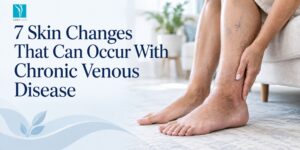
Chronic venous disease can affect both skin colour and texture. The seven changes worth recognising are brown pigmentation, venous eczema, redness or inflammation, shiny or fragile skin, lipodermatosclerosis, atrophie blanche and skin breakdown that can progress to a venous ulcer.
1. Brown, Rust-Coloured or Reddish Pigmentation
This is one of the most recognised pigmentation patterns linked with chronic venous disease. It commonly appears around the ankle or lower calf rather than uniformly across the whole leg.
With sustained venous pressure, red blood cells and their breakdown products can escape into surrounding tissue. Iron-containing pigment called haemosiderin can then contribute to a brown or rusty appearance.
A typical illustrative scenario is someone who has had ankle swelling after long days standing for several years and gradually notices a brown patch developing above the inner ankle. That pattern is worth assessing rather than treating only as a cosmetic stain.
2. Venous Eczema or Stasis Dermatitis
Venous eczema can look like ordinary dermatitis: red, itchy, dry, flaky or scaly skin. In some people it can become inflamed, cracked or weepy.
The important clue is context. If the rash sits on the lower leg and is accompanied by swelling, aching, heaviness or visible varicose veins, venous disease becomes one of the possible contributors.
The Australian clinical literature describes venous eczema as a manifestation of established venous disease associated with sustained venous hypertension. It can occur with or without a venous ulcer.
3. Redness, Inflammation and Irritated Skin
Chronic venous pressure can promote inflammation in the tissues. The skin may look redder than usual, feel irritated or become more sensitive.
However, redness is not specific to veins. Infection, dermatitis, allergic reactions, trauma and a blood clot can also produce redness. New redness that is painful, hot, rapidly spreading or associated with significant swelling should not be assumed to be ordinary venous eczema.
4. Shiny, Tight or Fragile-Looking Skin
Persistent ankle or lower-leg swelling can stretch the skin, making it appear shiny or tight. Chronic inflammation may also make the surface more delicate, dry or prone to cracking.
This change can be subtle. A person may first notice that socks leave deeper marks, the ankle looks puffier at the end of the day and the skin feels tighter than it used to. Those symptoms are more informative when considered together than when judged one at a time.
5. Thickening and Hardening Called Lipodermatosclerosis
Lipodermatosclerosis describes inflammation and scarring in the skin and fat of the lower leg. The tissue can become thick, firm and “woody”, sometimes with increased pigmentation.
Because lipodermatosclerosis can reflect more established venous disease, it is not a change to manage with moisturiser alone. The skin finding may reflect more established chronic venous disease and warrants clinical assessment.
6. White Scar-Like Areas Called Atrophie Blanche
Atrophie blanche appears as small white or pale scar-like patches, often around the lower leg or ankle. It can occur in chronic venous disease, sometimes in areas where the skin has previously ulcerated.
It is important not to diagnose it from a photo. White scar-like patches can have more than one cause, so a clinician may need to consider venous disease alongside other possible explanations.
7. Skin Breakdown and Venous Ulceration
The most concerning progression is a wound that breaks through the skin and does not heal normally. Venous ulcers often occur around the lower leg or ankle and can be associated with swelling, pigmentation, eczema, lipodermatosclerosis or atrophie blanche.
Venous ulcers can occur alongside swelling, pigmentation, eczema and other signs of chronic venous disease.
Read more about venous leg ulcers:
https://www.veincare.com.au/conditions/venous-leg-ulcers/
Why Does Chronic Venous Disease Change Skin Colour and Texture?
Skin changes develop when blood does not return efficiently through the leg veins and pressure remains elevated. Fluid and blood components can leak into surrounding tissue, triggering swelling, inflammation, iron-containing pigment deposits and, over time, fibrosis or breakdown of vulnerable skin.
People searching for varicose veins skin discolouration causes are often looking for one single explanation. In reality, the visible change is usually the result of several linked processes.
Venous Pressure Rises
Damaged or poorly functioning venous valves can allow reflux, meaning blood moves in the wrong direction. This increases pressure lower in the leg, especially while standing.
Fluid Moves Into Surrounding Tissue
Higher venous pressure can encourage fluid to move into nearby tissues. This contributes to ankle swelling and a feeling of tightness.
Red Blood Cells and Pigment Can Escape
Microscopic leakage of red blood cells into tissue can leave iron-containing haemosiderin behind after the cells break down. That is one reason venous pigmentation often looks brown or rust-coloured.
Ongoing Inflammation Changes Skin and Fat
When inflammation persists, the skin can become itchy, eczematous, thickened or fibrotic. In more advanced disease, lipodermatosclerosis and fragile skin may develop.
Skin Integrity Can Eventually Fail
Chronically inflamed, swollen or fibrotic tissue may become less resilient. Minor trauma can then be harder to recover from, increasing the risk of skin breakdown and ulceration.
According to Victoria’s statewide varicose-vein referral criteria, pigmentation, eczema, lipodermatosclerosis and atrophie blanche fall within the C4 clinical category of chronic venous disease. Healed and active venous ulcers are classified as C5 and C6 respectively.
External source:
https://www.health.vic.gov.au/statewide-referral-criteria/varicose-veins
How Can You Tell When Skin Changes May Be Vein-Related?
Vein-related skin changes are more likely when they affect the lower leg or ankle and occur with heaviness, aching, swelling or visible varicose veins. Appearance alone cannot confirm the cause, because several skin, inflammatory and circulation conditions can produce similar changes.
A vein-related pattern becomes more plausible when several features occur together:
- Pigmentation is concentrated around the ankle or “gaiter” area.
- Symptoms worsen after long periods of standing or sitting.
- Ankle swelling develops later in the day.
- The leg feels heavy, achy, tight or itchy.
- Visible varicose or reticular veins are present.
- The skin has gradually become firmer, drier or more fragile.
- There has been a previous DVT or previous vein treatment.
Read more about varicose vein symptoms:
https://www.veincare.com.au/varicose-veins/symptoms/
Read more about the causes of varicose veins:
https://www.veincare.com.au/varicose-veins/causes/
A Practical Pattern-Recognition Framework
Location: Venous skin changes frequently cluster around the lower calf and ankle.
Timing: Symptoms may be more noticeable after prolonged standing and less prominent after rest or elevation, although this pattern is not diagnostic.
Companion symptoms: Swelling, heaviness, aching, itching and visible veins make a venous cause more plausible.
Progression: A colour change that is spreading, becoming thicker, developing eczema or breaking down deserves more attention than a stable mark that has an obvious non-venous cause.
Alternative causes: Infection, arterial disease, dermatitis, trauma, medications, diabetes-related problems and other skin disorders can mimic or coexist with venous disease.
This is why a photo or online symptom list cannot replace a clinical assessment.
What Does a Varicose Veins Skin Discolouration Assessment Involve in Australia?
Assessment starts with your symptom history and physical examination, then may include duplex ultrasound when venous reflux is suspected. The aim is to determine whether the skin change is vein-related, map the underlying vein pattern and identify other possible causes.
A useful way to think about the appointment is that the clinician is not simply “looking at the colour”. The assessment connects the skin finding with circulation, vein anatomy and your medical history.
- Describe the change precisely: Note when it began, whether it is spreading, whether it itches or hurts, and whether swelling changes during the day; the timeline helps distinguish chronic progression from an acute problem.
- Examine the leg and skin: The clinician looks for visible veins, oedema, tenderness, eczema, pigmentation, hardening, ulcers and asymmetry; these findings help build a clinical picture rather than relying on one symptom.
- Use duplex ultrasound when indicated: Ultrasound can assess blood flow, identify reflux and map veins below the surface; this matters because the visible vein or skin change may be connected to a deeper abnormal vein.
- Discuss the findings before treatment: The clinician explains whether monitoring, skin care, compression, further investigation or a vein procedure may be appropriate; treatment should follow the diagnosis, not precede it.
Learn more about vascular ultrasound:
https://www.veincare.com.au/vascular-ultrasounds/
See what to expect at your first vein clinic consultation:
https://www.veincare.com.au/what-to-expect-from-your-vein-clinic-consultation/
What Treatment Questions Should You Ask if Skin Changes Are Present?
Treatment should be based on the underlying diagnosis rather than skin colour alone. Ask whether venous reflux is present, whether the skin may improve, whether compression is suitable, which treatment fits your vein anatomy, and what skin care or follow-up is needed.
Useful varicose veins skin discolouration treatment questions include:
- Is the pigmentation actually related to chronic venous disease?
- Is there reflux on duplex ultrasound?
- Are there signs of venous eczema, lipodermatosclerosis or ulcer risk?
- Should another skin or vascular condition be ruled out?
- Is compression appropriate for me, and what strength should be used?
- If a procedure is recommended, which abnormal vein is being targeted and why?
- Is the goal symptom control, prevention of progression, cosmetic improvement or a combination?
- How much of the existing pigmentation is likely to remain?
- What should I do if the skin becomes more painful, red, swollen or begins to break down?
Treatment of underlying venous disease does not guarantee that established pigmentation or scarring will completely disappear. Some changes can persist even after the vein problem is addressed, which is why realistic expectations matter.
Vein Care’s varicose vein treatment page explains that treatment planning depends on symptoms, vein anatomy, medical history, ultrasound findings and whether reflux is present.
Options may include sclerotherapy, ultrasound-guided sclerotherapy, endovenous laser treatment, cyanoacrylate vein glue or ambulatory phlebectomy, depending on assessment.
When Should You See a Doctor About Lower-Leg Skin Changes?
Arrange medical assessment when lower-leg discolouration is new, persistent, spreading or accompanied by swelling, itching, pain, hardening, bleeding or a slow-healing wound. Seek urgent care for sudden one-sided swelling, severe pain, marked warmth or redness, significant bleeding, chest pain or breathlessness.
The search varicose veins skin discolouration when to see a doctor often leads people to focus only on colour. Timing should instead depend on the associated symptoms and how quickly they developed.
Timing Guide
| Situation | Suggested response |
| Stable brown pigmentation with long-standing varicose veins and no acute symptoms | Arrange a routine clinical assessment, particularly if the area is progressing |
| New eczema, increasing ankle swelling, firm skin or recurrent inflammation | Arrange assessment sooner rather than relying only on self-care |
| Slow-healing wound or skin breakdown near the ankle | Seek medical assessment promptly |
| Significant bleeding from a varicose vein | Seek urgent medical care |
| Sudden one-sided leg swelling, pain, redness or warmth | Seek immediate medical assessment for possible DVT or another acute cause |
| Chest pain, sudden breathlessness, coughing blood, fainting or severe breathing difficulty | Call Triple Zero (000) for emergency care |
According to Healthdirect, you should see a doctor immediately for signs of DVT such as a red, swollen leg and call Triple Zero (000) for chest pain or trouble breathing.
External source:
https://www.healthdirect.gov.au/deep-vein-thrombosis
Vein Care also provides information on deep vein thrombosis:
https://www.veincare.com.au/conditions/deep-vein-thrombosis/
What Can You Do While Waiting for Assessment?
While waiting for assessment, protect the skin, move regularly and avoid treating a new rash or discolouration with strong creams or compression unless suitability is known. Gentle skin care and symptom tracking may help, but they cannot diagnose or correct venous reflux.
Use a plain moisturiser on intact dry skin: Reducing dryness can help protect the skin barrier, but stop and seek advice if a product causes irritation.
Avoid scratching itchy areas: Repeated scratching can break fragile skin and create a wound that is harder to heal.
Keep moving during the day: Regular walking and calf movement support venous return and are generally preferable to remaining still for long periods.
Elevate your legs when comfortable: Some people find this reduces swelling or heaviness, but it does not replace assessment when skin changes are progressing.
Track changes with dated photos: Photographing the same area in similar lighting every one to two weeks can help you and your clinician see whether pigmentation, swelling or skin breakdown is progressing.
Do not start strong topical medicines on your own: Redness or itch may have several causes, and inappropriate treatment can mask infection or irritate already vulnerable skin.
Do not assume compression is suitable: Medical compression can be helpful in venous disease, but arterial circulation and other factors may need to be considered before the correct type and pressure are chosen.
What Should Australians Know About Costs, Medicare and Treatment Planning?
In Australia, vein-care costs depend on the consultation, ultrasound, procedure, number of sessions and Medicare eligibility. Some procedures may attract a Medicare benefit when clinical criteria are met, while cosmetic treatment generally does not, so ask for a personalised estimate.
For varicose veins skin discolouration Australia searches, it is important to separate a Medicare schedule fee from the amount a private clinic charges.
The Medicare Benefits Schedule lists discolouration among the significant symptoms that may support eligibility for certain varicose-vein procedure items when the other clinical requirements are met.
For example, item 32520 requires duplex-documented reflux of at least 0.5 seconds in the relevant saphenous vein and has a schedule fee of A$638.70, as updated on 1 July 2026. This is a schedule fee, not a guaranteed clinic price or out-of-pocket cost.
External source:
https://www9.health.gov.au/mbs/fullDisplay.cfm?q=32520&type=item
At Vein Care, treatment costs depend on the assessment findings, whether ultrasound is required, the treatment type, number of veins or sessions and Medicare eligibility. Ask for a written personalised estimate before committing to a procedure.
Conclusion
Persistent lower-leg pigmentation can be a visible sign of chronic venous pressure, particularly when brown staining occurs with swelling, eczema-like irritation, hardening or a slow-healing wound. The skin change alone does not confirm the diagnosis, so assessment should focus on the whole clinical pattern.
If you have persistent or progressing lower-leg skin changes, consider booking a vein assessment with Vein Care in Melbourne: https://www.veincare.com.au/
The appropriate next step monitoring, skin care, ultrasound, compression or treatment should be based on clinical findings rather than appearance alone.
FAQ'S
Can Skin Discolouration Happen Without Obvious Varicose Veins?
Yes, visible bulging veins are not required for venous disease to be present. Abnormal reflux can occur in veins beneath the skin, while the first obvious signs may be ankle swelling, itching or pigmentation. Clinical examination and duplex ultrasound, when indicated, can help determine whether an underlying venous pattern is contributing.
Can Chronic Venous Disease Affect Only One Leg?
Yes, venous disease can be more pronounced in one leg, depending on previous clots, vein anatomy, injury, pregnancy-related changes or where reflux develops. One-sided symptoms still need proper assessment, particularly if swelling or colour change is new. Sudden unilateral swelling, pain or redness should be assessed urgently.
Does Brown Skin Around the Ankle Always Mean a Vein Problem?
No, brown or reddish pigmentation has several possible causes, including inflammatory skin conditions, previous injury, infection and other circulation problems. Venous disease becomes more likely when pigmentation sits around the ankle or lower calf and occurs with swelling, heaviness, aching, eczema-like irritation or visible varicose veins.
Will Moisturiser Remove Venous Pigmentation?
No, moisturiser can help dry, itchy or fragile skin, but it does not remove the venous pressure or pigment already deposited in tissue. If the discolouration is related to chronic venous disease, management needs to address both skin care and the underlying circulation problem identified during clinical assessment.
Are Compression Stockings Safe for Everyone With Discoloured Legs?
No, compression is useful for some venous conditions, but it is not automatically suitable for every person with leg discolouration or swelling. Arterial disease and other conditions can change what level of compression is safe. Use properly fitted medical compression only after a clinician has assessed whether it is appropriate for you.
Can a Previous DVT Cause Skin Changes Years Later?
Yes, some people develop post-thrombotic syndrome after a deep vein thrombosis, with ongoing swelling, discomfort, skin discolouration or ulcers in the affected leg. Healthdirect lists these as possible longer-term effects after DVT. Persistent symptoms should be discussed with a doctor rather than assumed to be ordinary varicose veins.